
Medically reviewed by Nachiket J. Patel, MD, FACC, FSCAI. Last medically reviewed: August 2026.
The names sound similar, and both tests may happen during the same cardiology visit. They do not show the same thing.
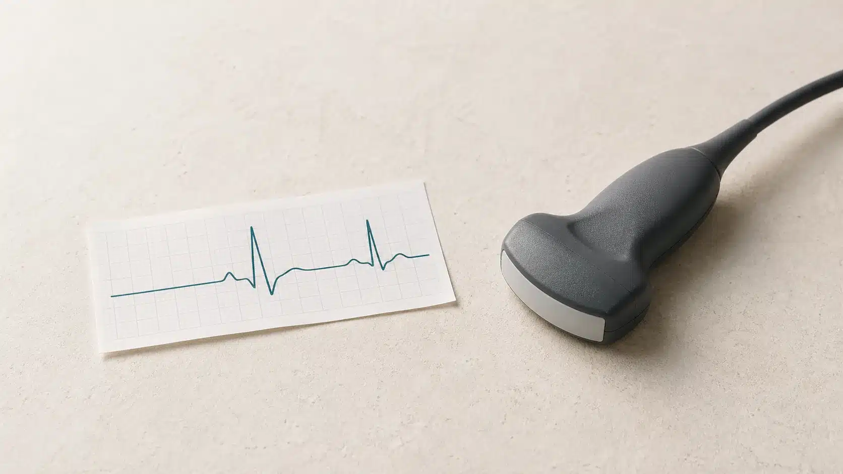
An electrocardiogram—abbreviated ECG or EKG—records the heart's electrical activity. An echocardiogram, often called an echo, uses ultrasound to make moving pictures of the heart. One is mainly an electrical test. The other is mainly a structure-and-function test.
The right question is not which test is “better.” It is which test can answer the clinical question in front of us.
What an ECG or EKG records
Small adhesive electrodes are placed on the chest and limbs. The machine records electrical signals as they travel through the heart; it does not send electricity into the body.
A standard ECG can help show:
- Heart rate and rhythm at the time of the recording
- Whether electrical signals are delayed or taking an unusual pathway
- Patterns that may accompany heart-muscle strain, a previous heart attack, or an active heart problem
- Electrical effects of certain medications or electrolyte abnormalities
The test is quick, painless, and valuable. It is also a brief snapshot. A normal ECG obtained while a patient feels well may not capture an intermittent rhythm problem that occurs once a week. In that situation, an ambulatory heart monitor may be more useful.
An ECG does not directly show how a valve opens, how large a heart chamber is, or how strongly the heart is pumping.
What an echocardiogram shows
For a standard transthoracic echocardiogram, a sonographer moves an ultrasound probe across several areas of the chest. Sound waves create moving images and Doppler measurements of blood flow.
An echo can help assess:
- The size, shape, and movement of the heart's chambers
- Overall and regional pumping function
- Whether a valve is narrowed or leaking
- Pressure clues within the heart and lung circulation
- Fluid around the heart
- Selected congenital abnormalities, masses, or blood clots
An echo is the main imaging test for many valve and heart-failure questions. But it does not provide a direct map of the inside of the coronary arteries. A normal resting echo therefore does not prove that there is no coronary plaque or explain every episode of chest discomfort.
The reported ejection fraction is useful, but it is not a complete score of heart health. Symptoms, valve function, wall thickness, filling, rhythm, blood pressure, and the clinical history still matter.
Why a clinician may order both
Consider a patient with shortness of breath and palpitations. The ECG may identify atrial fibrillation or another electrical pattern. The echo may show whether the chambers are enlarged, the valves are working normally, or pumping function has changed. The results complement each other.
Both tests may also be appropriate when evaluating:
- A new heart murmur
- Fainting or near-fainting
- Suspected heart failure
- Chest symptoms with an abnormal examination or ECG
- A known rhythm disorder that may be affecting heart function
- Follow-up of established heart valve disease
Not every patient needs both. Repeating an echo simply because time has passed may add little if the result is unlikely to change treatment. Conversely, a new symptom or examination finding may make repeat imaging important.
When the standard tests are not enough
“Echo” can refer to several different examinations. A transesophageal echocardiogram places a specialized probe in the esophagus to obtain closer views of selected structures. A stress echo compares heart-wall motion at rest and under stress. These tests have different preparation, risks, and purposes from a routine transthoracic echo.
Likewise, a standard ECG differs from a Holter monitor, patch monitor, telemetry monitor, or exercise ECG. The duration and setting should match the symptom pattern.
Other tests—such as cardiac CT, MRI, stress imaging, blood work, or coronary angiography—may be more appropriate when the question concerns coronary anatomy, inflammation, tissue characteristics, or blood flow during exertion. Good testing is not a collection. Each study should clarify a diagnosis, change risk assessment, or influence the plan.
What to tell the clinical team
Before either test, describe what prompted it in concrete terms. Helpful details include:
- What the symptom feels like
- Whether it begins at rest, during exertion, or after standing
- How long it lasts and how often it occurs
- Whether it comes with breathlessness, chest pressure, sweating, dizziness, or fainting
- Which medications and supplements you take
- Whether close relatives developed heart disease at an unusually young age
Bring prior ECGs and echo reports when possible. Comparison with an earlier study can be more informative than reading one result in isolation.
When not to wait for an outpatient test
Call 911 for new or severe chest pressure, severe shortness of breath, fainting with ongoing symptoms, sudden weakness or numbness on one side, difficulty speaking, or another possible heart-attack or stroke symptom. Do not drive yourself and do not wait for a scheduled ECG or echocardiogram.
An outpatient test is useful only after the immediate level of risk has been addressed.
Sources and Further Reading
- American Heart Association. Electrocardiogram (EKG or ECG). Last reviewed February 24, 2025.
- American Heart Association. Echocardiogram (Echo). Last reviewed February 24, 2025.
- American Heart Association. Key Patient Messages: 2025 Acute Coronary Syndromes Guideline.
- Otto CM, Nishimura RA, Bonow RO, et al. 2020 ACC/AHA Guideline for the Management of Patients With Valvular Heart Disease: Executive Summary. Circulation. 2021;143:e35–e71.
Related heart and vascular information
- Echocardiogram
- Ambulatory Heart Monitoring
- Palpitations and Arrhythmias
- Heart Valve Disease
- Shortness of Breath
Schedule an appointment
If you would like a focused review of your symptoms, risk, testing, or treatment options, request an appointment with Flow Heart & Vascular in Mesa, Arizona.
This article provides general education and does not replace individualized medical advice, diagnosis, or emergency care. Do not start, stop, or change medication or delay emergency care based only on information on this website.
